

 Implantable defibrillators and pacemakers have been around since the 1970s, but advances in materials science and 3-D visualization are transforming them from cumbersome life-support tools into streamlined therapies that could be props from Iron Man.
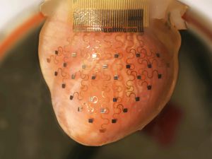
Professors John Rogers of the University of Illinois at Urbana-Champaign and Igor Efimov of Washington University in St. Louis have developed a new cardiac intervention that uses MRI and CT machines to scan a patient’s heart, 3-D printing a model from that data, and using the print to make a metallic mesh sleeve that can be implanted in the patient’s chest. The result looks like a gold doily and wraps around the heart to detect arrhythmias, deliver corrective electric shocks, and ultimately save lives.
Today, top-of-the-line implantable defibrillators determine if a patient needs a shock by parsing data collected from two or three electrodes. Efimov and Roger’s solution has over 30. These well-spaced contact points, paired with algorithms to detect cardiac problems, provide a high-definition view of the heart’s activity and apply more finely targeted adjustments.
Pairing these sensors with smartphones provides doctors and patients with a real-time data feed from the heart. Such a data stream could only be approximated in labs using millions of dollars of equipment a decade ago, but will transform the way care is delivered.
We’ve all seen medical dramas in which a doctor rips open a patient’s shirt, slaps a pair of defibrillator pads on the guy’s chest while yelling, “Clear!” and brings the guy back to life. If Rogers and Efimov have their way, that cliché will be replaced with an app.
SOURCE: Wired.com